Negative Staining
Negative Staining NoroVirus virus-like particles (VLPs) - Uranyless
New focus for application staining negative Virus.
Thanks to our feedback from UranyLess users around the world.
some users for specific applications reported that their results for viruses with negative staining were average to poor.
Our Laboratory Research and Development, has just made technical adjustments of the Uranyless which gives excellent results for all applications in positive or negative staining (We are always interested in your feedback, contact us at info @ deltamicroscopies. com).
We have developed for this application with a German Laboratory (especially thanking Dr André DIESSNER)
Icon Genetics GmbH – D – 06120 Halle / Saale.
we present you, their negative staining results : Novorirus
Image description: Electron micrographs of highly purified plant-produced norovirus virus-like particles (VLPs) examined by transmission electron microscopy (Zeiss EM900, Carl Zeiss Microscopy, Jena, Germany). Samples were collected on formvar coated cooper grids and contrasted with UranyLess. Micrographs were taken with a SM-1k-120 Variospeed slow scan camera (TRS, Moorenweis, Germany) using the iTEM software from Olympus SIS (Münster, Germany).
Important technical note:
the pH of the contrast reagent, plays an important role during your negative staining, always predict a more or less neutral pH, the acidity disrupts the spread of your particles (viruses, bacteria, liposomes, …). Uranyless pH 6,8 , uranyle acetate pH 4
During the preparation of your sample (Production, purification, …) there is sometimes sucrose, EDTA, buffers and others. the uranyless can react strongly with these additions and the contrast is too strong, in this case wash with distilled water just after spreading your sample drop on the film support of the grid and contrast afterwards.
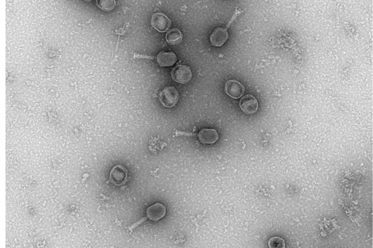
T6 Phage-Uranyless
T6 Phage
Sample preparation according to the following protocol:
- étalement de phage T6 sur une grille G300-Cu recouverte d’un film formvar carbone. Ionisation 1 mn.
Photos taken with a Hitachi HT7700 Transmission Electron Microscope by Nacer BENMERADI (R&D – Delta Microscopies – France).

Navigation
Contact
22b route de Saint Ybars 31190
Mauressac France
+(33) 5 61 73 60 14
info@deltamicroscopies.com


