Produit Uranyless Aqueous

Uranyless

Uranyless Aqueous

Uranyless aqueous is a contrasting solution that substitutes for uranyl acetate. As recommended by Reynolds (1963), it is highly advisable to reinforce the initial contrast (in our case, UranyLess) with lead citrate in a NaOH-saturated atmosphere to prevent contamination from atmospheric CO2.

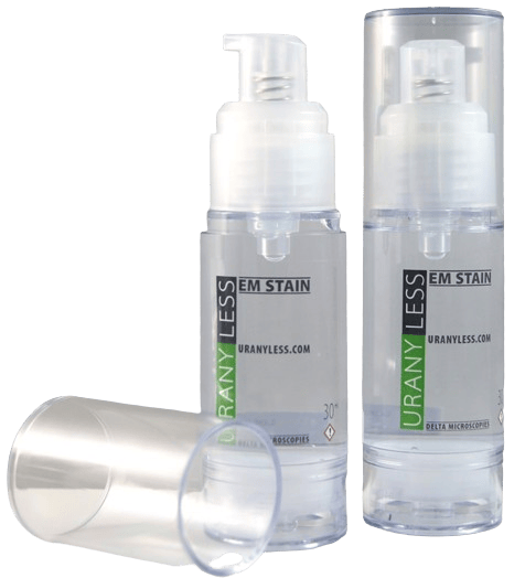
To facilitate the use of lead citrate during contrasting, it is packaged in AirLess bottles (without air and thus without CO2). You can store this 30 ml bottle for a long time without the risk of CO2 contamination, a common problem for microscopists.

The 30 ml AirLess bottle packaging allows you to contrast over 1500 grids.

This packaging is doubly effective: it provides long shelf life and generates minimal waste.

For using UranyLess aqueous in automated contrast devices like Leica’s EM Stain, UranyLess is available in a 200 ml bottle. It is worth noting that UranyLess aqueous is packaged in an AirLess bottle to facilitate drop dispensing only, as it is not affected by air or light.                                        

Uranyless aqueous bottle

Yeast

Preparation of the sample according to the following protocol:

  • Standard fixation with Glutaraldehyde – Osmium – Embedding in Epon.
  • Contrast with UranyLess followed by Lead Citrate.

Here are some results from Jeannine Lherminier (INRA – Dijon).

Photographs taken using Transmission Electron Microscopy

Trematodes

We present some results from Yann Quilichini (Microscopy Platform at the University of Corsica – Corte).

Sample preparation according to the following protocol:

  • Standard fixation with glutaraldehyde, osmium, embedding in Spurr resin.
  • Thin sections – contrast with aqueous UranyLess followed by lead citrate (according to Reynolds).

Photographs taken using Transmission Electron Microscopy.

Polymersomes-Uranyless

The IMRCP Laboratory in Toulouse, led by Anne-Françoise Mingotaud, tested UranyLess in comparison to uranyl acetate, which is at an acidic pH of 4 (seems to disrupt the molecular structure organization). They also compared observations using Cryo-SEM (scanning electron microscopy).

The chemical structure is organized as follows:

Observation using Scanning Electron Microscopy in Cryo mode.

Reconstructed Epidermis

We present the results from Audrey Houcine (CMEAB Toulouse).

Sample preparation according to the following protocol:

  • Standard fixation with glutaraldehyde, osmium, Epon/Araldite
  • Ultrathin sectioning, double contrast with Uranyless followed by lead citrate

Photographs taken using Hitachi HT7700 Transmission Electron Microscopy by Audrey Houcine.

Epiderme-reconstitué-5-973x1024 (1)
Glomerular Zone – podocytes – stalks – Kidney.

Muscle-Nerve-Mince

Sample preparation according to the following protocol:

  • Standard fixation with glutaraldehyde, osmium, Epon
  • Ultrathin sectioning, double contrast with Uranyless 1min followed by lead citrate 1min
Delta-muscle-cellules-23-953x1024 (1)
Longitunidal section of Mouse Skeletal Muscle- Nerve cup (dense area myelin sheath). Photo Nacer Benmeradi (R&D- Deltamicroscopies-France)
Delta-muscle00-1024x985 (1)
Detail View of Myocutes. Photo Nacer Benmeradi(R&D- DeltaMicroscopies-France)
Delta-muscle-squelettique-ct-3-1024x842
Cross section og Muscle Fibers- Mitochondria. Photo: Nacer Benmeradi (R&D- DeltaMicroscopies-France)
Delta-muscle-squelettique-ct-4-1024x1003
Myocyte. Photo: Nacer Benlmeradi (R&D- DeltaMicroscopies-France)

Mouse Ovarian Follicle

Sample preparation according to the following protocol:

  • Standard fixation with glutaraldehyde Fixative PFA, osmium, Epon
  • Ultrathin sectioning, double contrast with Uranyless 1min followed by lead citrate 1min
theque-interne
Theca Interna Mouse Ovarian Follicle. Photo: Nacer Benmeradi (R&D- DeltaMicroscopies-France)
theque-interne-2
Mitochondria in Typical Finger Glove Syeroid Synthesis in Cells (internal Thèque Ovarian Follicle). Photo: Nacer Benmeradi (R&D- DeltaMicroscopies-France)
Delta-cellule-folliculaire
Follicular Cell of the Corona Radiata a Mouse Ovarian Follicle. Photo: Nacer Benmeradi (R&D- DeltaMicrooscopies- France)
theque-externe1 (1)
Cell of the External Library of Mouse Ovarian Follicle. Photo: Nacer Benmeradi (R&D - DeltaMicroscopies-France)

Mouse Kidney

Mouse ovarian follicle

Sample preparation according to the following protocol:

  • Standard fixation with glutaraldehyde Fixative PFA, osmium, Epon
  • Ultrathin sectioning, double contrast with Uranyless 1min followed by lead citrate 1min
theque-interne
Theca Interna Mouse Ovarian Follicle. Photo: Nacer Benmeradi (R&D- DeltaMicroscopies-France)
theque-interne-2
Mitochondria in Typical Finger Glove Syeroid Synthesis in Cells (internal Thèque Ovarian Follicle). Photo: Nacer Benmeradi (R&D- DeltaMicroscopies-France)
Delta-cellule-folliculaire
Follicular Cell of the Corona Radiata a Mouse Ovarian Follicle. Photo: Nacer Benmeradi (R&D- DeltaMicrooscopies- France)
theque-externe1 (1)
Cell of the External Library of Mouse Ovarian Follicle. Photo: Nacer Benmeradi (R&D - DeltaMicroscopies-France)

Kidney

Mouse Kidney

Sample preparation according to the following protocol:

  • Classic PFA- Glutaraldehyde, Osmium, Epon fixing.
  • Ultrafine section – 1 minute UranyLess contrast – 1 minute Lead Citrate.

Photos taken with a Hitachi HT7700 Transmission Electron Microscope by Nacer BENMERADI (R&D – Delta Microscopies – France).

Delta-rein4-1024x1014
Glomerular Zone – podocytes – stalks – Kidney.. Photo: Nacer Benmeradi (R&D- DeltaMicroscopies-France)
Delta-rein31-1024x946
Podocytes – stalks – Kidney. Photo: Nacer Benmeradi (R&D- DeltaMicroscopies-France)
Delta-Rein3-1024x1010
Mouse kidney. Nacer Benmeradi (R&D DeltaMicroscopies-France)
rein2-933x1024
Basal invaginations – hemi- desmosomes – basal blade: increase the exchange surface – Kidney. Nacer Benmeradi (R&D- DeltaMicroscopies-France)

Cardiac muscle - Uranyless

Mouse cardiac muscle

Preparation of the sample using following protocol:

  • classic Fixing Glutaraldehyde, Osmium, Epon Ultrafine
  • Cups, Double Uranyless Contrast and Lead Citrate
Image1-926x1024
Mouse Cardiac Muscle. Photo: Nacer Benmeradi (R&D- DeltaMicroscopies-France)
Muscle-cardiaque-souris-2-927x1024
Mouse Cardiac Muscle. Photo: Benmerdi Nacer (R&D- DeltaMicroscopies-France)
Image2-926x1024
Mouse Cardiac Muscle. Photo: nacer benmeradi (R&D- DeltaMicroscopies-France

Plant Tissu

Mouse cardiac muscle

Preparation of the sample using following protocol:

  • classic Fixing Glutaraldehyde, Osmium, inclued in epon
  • Double Uranyless Contrast and Lead Citrate
feuille-1-lherminier
Plant Leaf. Photo: Jeannine Lherminier
feuille-2-lherminier
Plant Leaf. Photo: Jeannine lherminier

Phage T6

Sample preparation according to the following protocol:

  • Staggering Phage t6 on a G300-Cu grid covered with formvar film. Lonization 1 min.
negative staining phage t6
Glomerular Zone – podocytes – stalks – Kidney.. Photo: Nacer Benmeradi (R&D- DeltaMicroscopies-France)
mouse kidnay negative staining
Podocytes – stalks – Kidney. Photo: Nacer Benmeradi (R&D- DeltaMicroscopies-France)
phage-T6-2 (1)
Mouse kidney. Nacer Benmeradi (R&D DeltaMicroscopies-France)
phage T6 negative staining
Basal invaginations – hemi- desmosomes – basal blade: increase the exchange surface – Kidney. Nacer Benmeradi (R&D- DeltaMicroscopies-France)

Cross sectional Bacteria/Bacteri E-Coli

Sample preparation according to the following protocol: Cross sectional Bacteria

  • Fixing Classic Glutaraldehyde Osmium, EPON
  • Cutting ultrafine, double contarst Uranyless and lead Citrat
C.-Longin
Cross sectional Bacteria. Photo: Christine Longia (INRA JOUy en Josas)
C.-Longin-1
Bacteria: Christine Longia (INRA Jouy en Josas)

Bacteri E-coli

  • Negative Staining for 2 minutes Uranyless bacteria like E-coli (adherent and invasive (ACSI) LF82) Which have Pili and Flagella .
bacterie negative staining
Basal invaginations – hemi- desmosomes – basal blade: increase the exchange surface – Kidney. Nacer Benmeradi (R&D- DeltaMicroscopies-France)

Sacculina crustacean/Intestine

Sample preparation according to the following protocol: Sacculina Crustace (small parasitic crustacean)

  • Fixing Classic Glutaraldehyde Osmium, EPON inclusion 
  • Fines Cups- Contrast to the aqueous Uranyless 60°C on a Hotplate without Lead Citrat post coloring
djediat-uranyless-980x1024
Sacculina cuticle area. Photo: Dedjat Chakib (Natural Museum Paris)

Intestine

  • Classic Glutaraldehyde Osmium, EPON inclusion 
  • Ultrafine cup-Contrat Uranyless and Lead Citrat
intestin-2
Basal invaginations – hemi- desmosomes – basal blade: increase the exchange surface – Kidney. Nacer Benmeradi (R&D- DeltaMicroscopies-France)

Liver Mouse and andrenal Gland

Sample preparation according to the following protocol: Liver Mouse and gherbil Sahara

  • Fixing Classic Glutaraldehyde Osmium, EPON
  • Cutting ultrafine, double contarst Uranyless and lead Citrat
Foie-hépatocyte-1-1021x1024
Hepatocyte-Perinuclear region. Photo: Nacer Benmeradi (R&D Deltamicroscopies France)

Andrenal Gland gherbil Sahara

  • Fixing Classic Glutaraldehyde Osmium, EPON
  • Cutting ultrafine, double contarst Uranyless and lead Citrat
cortico-surrénale-6
Andrenocortica. Photo: Nacer Benmeradi (R&D Deltamicroscopies, France)
cortico-surrénale2
Andrenocortica. Photo: Nacer Benmeradi (R&D Deltamicroscopies, France)

Cells in culture - Uranyless

Sample preparation according to the following protocol:

  • Classic PFA- Glutaraldehyde, Osmium, Epon fixing.
  • Ultrafine section – UranyLess contrast – Lead Citrate.

Photos taken with a Hitachi HT7700 Transmission Electron Microscope by Nacer BENMERADI (R&D – Delta Microscopies – France):

SEE ALSO

Contact

22b route de Saint Ybars 31190 

Mauressac France

+(33) 5 61 73 60 14

info@deltamicroscopies.com

Get Connected

Scroll to Top